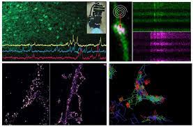
Progress in microscopy has a long history of triggering major advances in neuroscience, and it is accelerating on many fronts: morphological and functional labeling, microscope instrumentation and imaging modalities, image processing and data analysis etc. These innovations are potentiating our ability to monitor, measure and manipulate biological structures and activities inside complex and intact nervous system model systems with ever higher throughput, robustness, precision and sensitivity and on ever wider temporal and spatial scales.
This Cajal course will bring together leading developers and practitioners of cutting-edge imaging techniques that push the frontiers of neuroscience research. The course follows up on previous Cajal school editions on this topic. The course will cover a broad spectrum of concepts and practical techniques, both classic and new, to provide a solid basis and critical guidance for newcomers and experienced users alike, wishing to pick up skills and learn about new developments and avenues in microscopy and its impact on neuroscience.
The applications will span a wide spectrum of neurobiological topics and preparations in an exemplary fashion, from brain development, plasticity and neuro-immune system interactions in cell cultures, brain slices and in vivo using the mouse and zebrafish brain as main model systems.
A series of pedagogical lectures and seminars will be complemented by hands-on practical training in small groups using experimental setups and tools provided by the Bordeaux School of Neuroscience, the Bordeaux Imaging Center, the course faculty & instructors, including a number of leading research labs on Bordeaux Neurocampus, as well as brand-new demo equipment from microscope manufacturers.
Course directors
- Valentin Nägerl (Bordeaux University, France)
- Francesca Odoardi (Göttingen University, Germany)
- Jan Huisken (Göttingen University, Germany)
September 30 – 11:00am
Alexander Flügel University Medical Center Göttingen, Germany
Intravital 2-photon microscopy of CNS autoimmunity.
October 1 – 9:00am
Anna-Sophia Wahl LMU Munich, Germany
High-resolution 2photon imaging in-vivo to unveil key principles of neuronal repair.
October 1 – 11:00am
Dmitri Rusakov, University College, UK
Principles and neuroscience applications of time-resolved fluorescence microscopy.
October 2 – 9:00am
Florian Engert, Havard University, USA
Dissection of a zebrafish integrator circuit through correlated light and electron microscopy.
October 2 – 11:00am
Moritz Helmstaedter, MPI for Brain Research, Germany
Cerebral Cortex Connectomics.
October 5 – 11:00am
Jan Huisken, Georg-August University Göttingen, Germany
Flamingo: Fast and gentle volumetric imaging inside and outside the optics lab.
October 10 – 9:00am
Christophe Zimmer, Institut Pasteur, France
Deep learning: principles and applications to biomedical imaging.
October 12 – 9:00am
Francesca Odoardi, Georg-August University Göttingen, Germany
Intravital imaging in neuroimmunology: limitations and challenges.
October 14 – 9:00am
Johann Danzl, Institute of Science and Technology Austria, Austria
Reconstructing brain tissue with light microscopy.
October 15 – 9:00am
Laurent Groc, Bordeaux University, France
Using single molecule imaging to unveil membrane protein organization and extracellular space in the brain.
October 15 – 11:00am
Hans-Ulrich Dodt, Medical University of Vienna, Austria
From mice to man – new results of ultramicroscopy of cleared sample.
October 17 – 9:00am
Valentin Närgel, Bordeaux University, France
STED imaging of living brain microstructures.
I am text block. Click edit button to change this text. Lorem ipsum dolor sit amet, consectetur adipiscing elit. Ut elit tellus, luctus nec ullamcorper mattis, pulvinar dapibus leo.