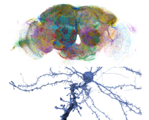
The biological factors shaping the synaptic connectivity of neuronal circuits are complex and multifaceted, depending on cell types, functional activity, homeostasis, and more. Mapping brain wiring at the level of both local circuits and across brain-wide projections is a key aspect of understanding how nervous systems develop, learn, process information and generate behaviour. Recent advances in molecular biology, tissue processing, computational methods, and microscopy have enabled a revolution in understanding structural connectivity with cellular and synaptic resolution. Large-scale electron microscopy volumes provide nanometer-scale maps of anatomy and connectivity of whole invertebrate brains and millimetre-scale regions of vertebrate brains, while light-microscopic methods can highlight genetically defined connections and enable brain-wide reconstruction of neurons. Together, these complementary approaches yield powerful insight into the neuroanatomy and connectivity of the nervous system with single-cell resolution.
This course will provide students with a broad introduction to contemporary methods of studying neuronal connectivity with lectures from experts in the field. It also provides practical project-based instruction in experimental methods of circuit tracing and reconstruction with light microscopy (light sheet or 2-photon), as well as the computational analysis of rich electron microscopy connectomes in flies and mice. Students will consider the strengths and limitations of different techniques and how they can be used to address key problems in circuit neuroscience.
Course director & co-directors
- Gregory Jefferis (MRC LMB and Cambridge University, UK)
- Jinny Kim (Korea Institute of Science and Technology, Korea)
- Nicolas Renier (Paris Brain Institute, France)
- Casey Schneider-Mizell (Allen Institute of Brain Science, USA)
September 19 – 9:00am Valentin Naegerl (Bordeaux University, France)
STED imaging of brain microanatomy
September 21 – 9:00am Claire Wyart (ICM Institute for Brain and Spinal Cord, France)
Optical methods to probe Connectivity of sensorimotor circuits in brainstem and spinal cord.
September 23 – 9:00am Jae-Byum Chang (Korea Advanced Institute of Science and Technology, KAIST, Korea)
Super-resolution imaging of whole mouse embryos via whole-body expansion microscopy.
September 25 – 9:00am Alexandra Pacureanu (European Synchrotron Radiation Facility, France)
September 26 – 9:00am Jonny Kohl (Francis Crick Institute, UK)
Form, function and flexibility of parenting circuits.
September 28 – 9:00am Moritz Helmstaedter (Max Planck Institute for Brain Research, Germany)
Cerebral Cortex Connectomics.
September 29 – 9:00am Hiroki Ueda (UTokyo/RIKEN BDR, Japan)
Towards Human Systems Biology of Sleep/Wake Cycles: Phosphorylation Hypothesis of Sleep.
October 2 – 9:00am Constantin Pape (Georg August Universität Göttingen, Germany)
Instance Segmentation Methods for Large Volumetric EM.
October 3 – 9:00am Christel Genoud (University of Lausanne, Switzerland)
VolumeEM techniques to study the central nervous system.
October 3 – 11:00am Casey Schneider-Mizell (Allen Institute of Brain Science (USA)
Martin Carbo Tano – Paris Brain Institute, France
Andrew Champion – Cambridge University, UK
In Cho – Korea Advanced Institute of Science and Technology, Korea
Yulia Dembitskaia –Bordeaux University, France
Kathi Eichler – Leipzig University, Germany
Leila Elabbady – University of Washington, USA
Jordan Girard –Bordeaux University, France
Jihyun Kim– Korea University, Korea
Sahil Loomba – Max Planck Institute for Brain Research, Germany
Philipp Schlegel – University of Cambridge, UK
Thomas Topilko – Gubra, Danemark
Alba Vieites Prado – Universitad de Santiago de Compostela, Spain
Fabian Voigt – Harvard University, USA
Yagmur Yener – Max Planck Institute for Brain Research, Germany
Light Microscopy and functional, molecular methods:
– Choice of labelling strategy: use of specific cre lines (for instance, the GENSAT project); finding specific markers for cell populations; using viral vectors and intersectional genetics (Dual or triple injections, transsynaptic tracing, Tango system, mGRASP, etc.).
– Tissue preparation for imaging: tissue clearing: choice of methods, considerations for the resolution needed and type of molecular labelling (Ueda, Renier); expansion microscopy methods: when to use them, and which iteration (Jae-Byum Chang)
– Imaging strategy: use of scanning microscopes: confocal or 2p, in intact samples or using serial sectioning; use of light sheet microscopy: commercial systems (eg. Miltenyi’s Blaze or Zeiss Z7), and custom systems (Mesospim).
– Analysis of imaging data: use of neuron mapping pipelines for whole brain data obtained from light sheet microscopy or from sections (eg. ClearMap, TrailMap, WholeBrain, etc). (Ueda, Renier); se of virtual reality-assisted tools for single neuron reconstructions from 3D datasets (eg. SyGlass, Vision4D…).
Electron microscopy synaptic connectomics:
At the end of this course, the students will be familiar with all of the steps that go into producing and analysing large scale, synaptic resolution EM connectomics datasets, summarised as below. Detailed analysis projects tailored by student interest will use public datasets and open source tools in which their directors and their colleagues are experts. These include the microns mouse cortical cubic millimetre dataset and fly CNS datasets including the hemibrain, flywire.
Keynote Speakers
Gwyneth Card – Columbia University, USA
Jae-Byum Chang – KAIST, South Korea
Christel Genoud – University of Lausanne, Switzerland
Moritz Hemlstaedter – Max Plank Institute for Brain Research, Germany
Johannes Kohl – The Crick Institute, UK
Valentin Nägerl – University of Bordeaux, France
Alexandra Pacureanu – European Synchrotron Radiation Facility, France
Constantin Pape – Georg August Universität Göttingen, Germany
Hiroki Ueda – Laboratory for Synthetic Biology, Japan
Claire Wyart – ICM Institute for Brain and Spinal Cord, France